~600 microns
field of view, 20x
Micro Solutions / Life Science
Visualization, photoactivation and electrophysiology on slices: up to 16 analog channels in one system
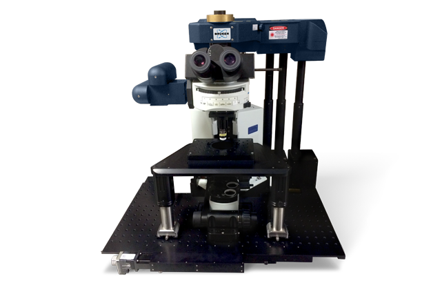
Visualization, photoactivation and electrophysiology on slices: up to 16 analog channels in one system
Multiphoton system based on Nikon, Olympus and Zeiss microscopes for slices and cultures
Multiphoton system for slices, cell cultures and combined optical and electrophysiological experiments.
Ultima In Vitro combines visualization, photoactivation and recording of electrical signals. The second scanning path, synchronization of external devices and Prairie View allow you to build complex protocols for neural networks and tissue explant.
Field of view is about 600×600 μm with a 20× lens. Raster scanning reaches 1.65 fps at 512×512, optionally 30 fps with a resonance scanner; optical input is optimized for 690–1300 nm.
field of view, 20x
512×512, resonance scanner
multiphoton
analogue inputs/outputs
| Method of scanning | Cambridge 6mm pair of electroplating, raster and spiral scanning |
|---|---|
| Field of vision | ~600 × 600 μm with a 20× lens |
| Speed (raster) | 1.65 fps at 512×512 >12 fps at 64×64 |
| Speed (spiral) | 6 fps at 512×512; ~30 fps at 64×64 |
| Ankajing (option) | second set of Cambridge 3 mm galvanometers |
| Speed shooting (option) | 8 kHz resonance galvanometer; 30 fps at 512×512; >1300 fps for ROI 512×8 |
| The reflected channel | 1-4 Hamamatsu Multi-Alkali, upgrade to GaAsP |
|---|---|
| The passing channel | 1-2 Hamamatsu Multi-Alkali |
| Dodt / passing light | one-way Hamamatsu |
| Multiphoton laser | 690-1300 nm |
|---|---|
| Laser safety class | Class 1 |
| Basic microscopes | Nikon Ti-E, Ni-E; Olympus BX51WI, BX61WI; Zeiss Axio Observer/Examiner |
| Piezofocus. | range of 150 μm, accuracy of 0.05 μm |
| Analog inputs/outputs | 16 |
|---|---|
| Software | Prairie View |
Second set of 3 mm galvanometers for photo manipulation
High-speed photography
Increased sensitivity of the reflected channel