1.4 mm
field of view, 16x
Micro Solutions / Life Science
Deep in vivo imaging with a wide field of 1.4 × 1.4 mm and the possibility of growth up to Ultima 2Pplus
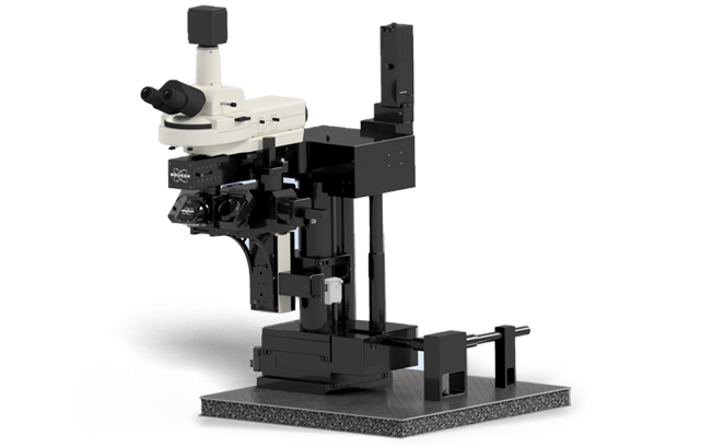
Deep in vivo imaging with a wide field of 1.4 × 1.4 mm and the possibility of growth up to Ultima 2Pplus
Basic multiphoton system with wide-angle signal collection and modular options
Modular multiphoton microscope with a large field of view for deep in vivo imaging of tissues.
The system is optimized for living objects and combines wide-angle signal collection with an expandable platform, and optogenetics, high-speed scanning, FLIM, multi-area photography and subsequent upgrade to Ultima 2Pplus are available.
The resonance scanner supports 30 fps at 512×512. For Z-positioning, a piezo drive is available with a step of 0.1 μm and a range of 150 μm; the motorized XY table has a stroke of 6 inches X and 3 inches Y.
field of view, 16x
512×512, resonance scanner
laser-entry
field-upgradation
large gap for working with animals
multi-pole editing and deep shooting
| Method of scanning | pair of 6 mm galvanometers |
|---|---|
| Field of vision (galvano) | 16×: 1.4×1.4 mm; 40×: 495×495 μm; 60×: 160×160 μm |
| Field of vision (resonance) | 16×: 982×982μm; 40×: 353×353μm |
| Raster speed (zoom 1x) | 2048×2048 – 0.334 fps; 1024×1024 – 0.787 fps; 512×512 – 1.572 fps; 64×64 – 12.329 fps |
| Time for the pixel | 0.4–100 μs in increments of 0.4 μs |
| Scanning setup | 1–2048 pix/line and rows/frames; zoom to 128; turn 360°; linear and point scans |
| Resonance scanner (option) | 8 kHz; 30 fps at 512×512 |
| Detectors. | 2 Multi-Alkali FEUs; optionally GaAsP Hamamatsu or 4 upper non-descanned detectors |
|---|---|
| Optical entrance | ultrashort pulse IR laser (2P or 3P), laser is not included |
| Epifluorescence | dichroic-mirror |
| Z-focus. | stroke of 25 mm, minimum pitch of 0.025 μm |
|---|---|
| Z-pieze | 0.1 μm step; range 400 or 1000 μm |
| Motorized base (option) | XY-movement ~19 mm (0.75 inches), step 0.3 μm |
| Motorized XY table | ≈152 × 76 mm (6 × 3 inches), step from 0.3 μm, accuracy 1 μm |
| Computer | specialized workstation |
|---|---|
| Software | Prairie View, PrairieLink |
| Imput/output | high-performance analog inputs/outputs, GPIO block, digital synchronization |
30 fps at 512×512, ROI with panning
Hypersensitivity
Visualization of the life time of fluorescence
Full photoactivation
Software power management of laser
Voltage-image
Off-axis tissue imaging
Translation to an inverted configuration