FN 28 mm
field
Micro Solutions / Life Science
FN 28mm and excitation up to 1700nm: imaging, photostimulation and electrophysiology in one experience
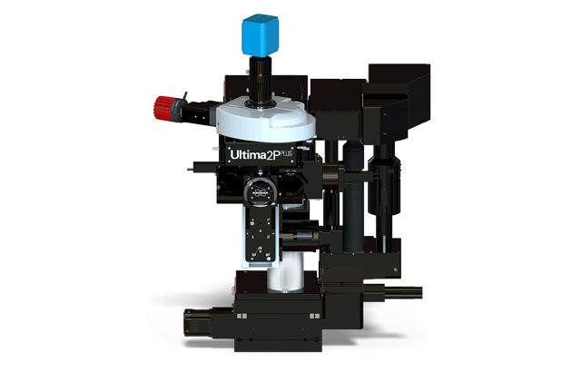
FN 28mm and excitation up to 1700nm: imaging, photostimulation and electrophysiology in one experience
Fully Optical Multiphoton Workstation for In vivo Research
Multiphoton workstation for deep imaging, photostimulation and electrophysiology in a single experiment.
Two independent scanning paths allow both image capture and photoactivation to occur simultaneously, and the motorized sample positioning and modular architecture support intravital protocols, optogenetics and high-speed volumetric photography.
The FN 28 mm optical field exceeds the standard FN 18 mm; the optical path is designed to excite up to 1700 nm for three-photon tasks. Optional resonance scanner provides up to 30 fps at 512 x 512.
field
three-photon
512×512, resonance scanner
phEU detectors
increased table gap for large animals
Photostimulation, FLIM, resonance scanner
| Method of scanning | Cambridge 6mm pair of electroplating, raster and spiral scanning |
|---|---|
| Field of vision | ~1.375 × 1.375 mm with a 16× lens (FN ≤ 28 mm) |
| Speed (raster) | 1.65 fps at 512×512; >12 fps at 64 px. |
| Speed (spiral) | 6 fps at 512×512; ~30 fps at 64×64 |
| Resonance scanner (option) | 8 kHz; ~30 fps at 512×512; >1300 fps for ROI 512×8 |
| Scanning setup | 1–2048 pix/line and rows/frame; zoom to 120×; 360° rotation; linear and point scans |
| Photomanipulation (option) | a second set of 3 or 6 mm galvanometers in the same scanning head |
| The Reflected Channel (non-descanned) | 1-4 Hamamatsu Multi-Alkali, upgrade to GaAsP |
|---|---|
| The passing channel (non-descanned) | 1–2 Hamamatsu Multi-Alkali FEU, upgrade to GaAsP |
| Others. | FEU for contrast Dodt and passing light; C-mount camera port |
| Multiphoton laser | 690-1700 nm |
|---|---|
| Epifluorescence | turret-powered |
| Visible lasers | fiber-entry |
| Laser safety class | Class 1 with a light-shielding casing |
| XY-table | ~15 cm X, ~7.5 cm Y, step ~0.3 μm |
|---|---|
| XY-base displacement | ~35 mm, step 0.1 μm |
| Z-focus. | 30 mm, step ~0.2 μm |
| Z-pieze | up to 1000 μm, step 0.05 μm |
| Software | Prairie View: Z- and T-series, Atlas montages, ROI, photoactivation, analog inputs/outputs for electrophysiology |
|---|
Simultaneous photostimulation or ankage during shooting
Holographic 3D Photo Stimulation of Groups of Cells
High-speed shooting 30 frames / s at 512×512
Optically corrected electrically adjustable lens, operating range of 450 μm
Visualization of the lifetime of fluorescence and phosphorescence
Introduce two lasers without rearrangement of wavelength
Full-field optogenetics
Off-axis shooting, optionally motorized
Moving a microscope or sample; Bridge Stage turns the system into a slicer
Fast volumetric shooting in synchronous with resonance scanner
Detection of weak signals in vitro and direct HVG (SHG)
Contrast of unpainted slices in electrophysiology
Translation to an inverted configuration